Friday, April 1, 2011

Super Solar Flare Predicition

I started looking at flare predication more closely with NASA's concern for a coming super solar flare..


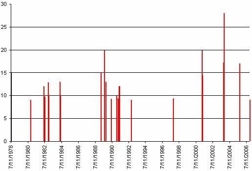
First I began with a listing of X-Class flares from 1975 forward…

Ranking   Day/Month/Year   X-Ray Class

----------------------------------------------
1                04/11/03              X28+
2                02/04/01              X20.0
2               16/08/89              X20.0
3               28/10/03              X17.2
4               07/09/05               X17
5               06/03/89              X15.0
5               11/07/78              X15.0
6               15/04/01              X14.4
7               24/04/84              X13.0
7               19/10/89              X13.0
8                15/12/82             X12.9
9                06/06/82             X12.0
9                01/06/91             X12.0
9               04/06/91              X12.0
9               06/06/91              X12.0
9               11/06/91              X12.0
9                15/06/91             X12.0
10              17/12/82             X10.1
10              20/05/84             X10.1
11              29/10/03             X10
11              25/01/91             X10.0
11              09/06/91             X10.0
12              09/07/82             X 9.8
12               29/09/89            X 9.8
13              22/03/91             X 9.4
13              06/11/97             X 9.4
14              24/05/90             X 9.3
15              05/12/06             X 9.0
15              06/11/80             X 9.0
15              02/11/92             X 9.0
Producing this graph;


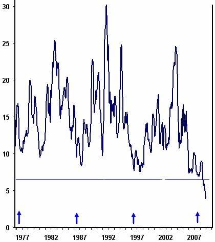
I then grabbed a five month averaged graph of the Ap Solar Progression for the same time period;


I overlaid these graphs with the six shortest solar cycles as I approximate them in length. It resulted in this…


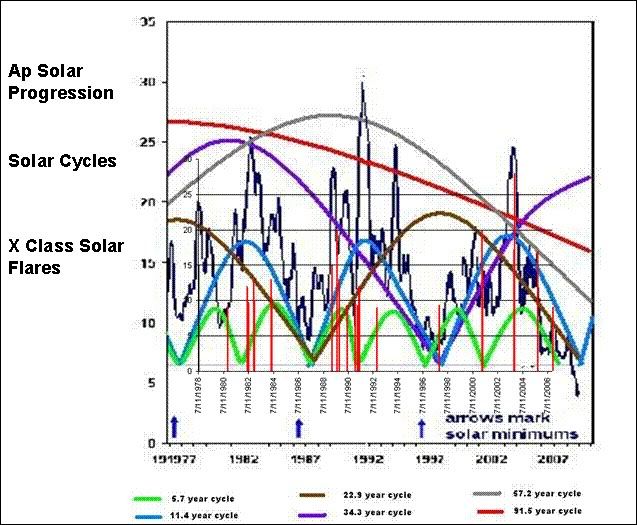
The April 11th 2003 flare (ranking #1 – ~X28) is a good example of how large flares occur when multiple solar cycles converge.


It also can be seen easily in 1982 and 1984.

The predominance of flares in ~1991 seem tied to the peak of the ~67 year solar cycle maxima.

Ranking    Day/Month/Year   X-Ray Class

----------------------------------------------
12              29/09/89             X 9.8
7                19/10/89             X13.0
14               24/05/90            X 9.3
11              25/01/91             X10.0
13              22/03/91             X 9.4
9                01/06/91             X12.0
9                 04/06/91            X12.0
9                 06/06/91            X12.0
9                 11/06/91            X12.0
9                 15/06/91            X12.0
11               09/06/91           X10.0
This just reinforces with me that solar flares are predictable when you correlate them to interactions between multiple solar cycles stumbling over each other.


So if we look to the last Gleissberg minima of ~1905/06 and looked for X Class solar flares we should see some reflection of flares that lie ahead in the next Gleissberg minima.


It would appear that the date of last Gleissberg minima was the autumnal equinox of 1906.


In pushing my solar cycle/s theory into the future for an approximation of when the next higher level convergence will occur…


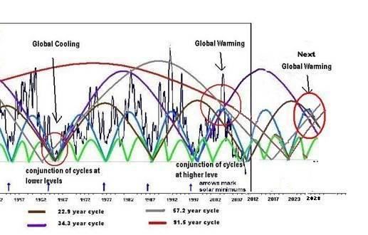
This is based on the Gleissberg minima occurring in solar cycle 25’s maxima

This should be prime time for solar flares.


This timing correlates with NASA’s window…

Long Range Solar Forecast
http://science.nasa.gov/science-news/science-at-nasa/2006/10may_longrange/

Solar flares appear predicatable as related to multiple solar cycles running concurrently and interacting with each other.


Friday, January 21, 2011

Melatonin, Magnetic Fields and Cancer

Since electricity, magnetic fields and grounding are all linked this may make sense.

As stated below melatonin blocks magnetic field induced DNA damage to cells, so melatonin reducing magnetophosphenes is totally feasible.

Melatonin and N-tert-butyl-α-phenylnitrone block 60-Hz magnetic magnetic field-induced DNA single and double strand breaks in rat brain cells

by H Lai - 1997 - Cited by 98 - Related articles

30 Jan 2007 ... Melatonin and N-tert-butyl-α-phenylnitrone block 60-Hz magnetic field-induced DNA single and double strand breaks in rat brain cells ...

ABSTRACT: In previous research, we have found an increase in DNA single-and double-strand breaks in brain cells of rats after acute exposure (two hours) to a sinusoidal 60-Hz magnetic field. The present experiment was carried out to investigate whether treatment with melatonin and the spin-trap compound N-tert-butyl- -phenylnitrone (PBN) could block the effect of magnetic fields on brain cell DNA. Rats were injected with melatonin (1 mg/kg, sc) or PBN (100 mg/kg, ip) immediately before and after two hours of exposure to a 60-Hz magnetic field at an intensity of 0.5 mT. We found that both drug treatments blocked the magnetic field-induced DNA single-and double-strand breaks in brain cells, as assayed by a microgel electrophoresis method. Since melatonin and PBN are efficient free radical scavengers, these data suggest that free radicals may play a role in magnetic field-induced DNA damage.
This study reveals that night adaptation reduces magnetophosphenes and melatonin is a dark adaptation;

Medical and Biological Engineering and Computing

Volume 18, Number 3, 326-334, DOI: 10.1007/BF02443387

Magnetophosphenes: a quantitative analysis of thresholds

P. Lövsund, P. Å. Öberg, S. E. G. Nilsson and T. Reuter

Low-frequency and transient magnetic fields of moderate flux densities are known to generate visual phenomena, so-called magnetophosphenes. In the present study, time-variable very low frequency (10–50 Hz) electromagnetic fields of moderate flux density (0–40 mT) were used to induce magnetophosphenes. The threshold values for these phosphenes were determined as a function of the frequency of the magnetic field both in normal subjects and colour defective ones. Maximum sensitivity occurred at a frequency of approximately 20–30 Hz, and with broad-spectrum light the threshold flux density was 10–12 mT. The threshold values were found to be dependent upon the intensity and the spectral distribution of the background light. Sensitivity decreased during dark adaptation. In certain respects deutans differed from subjects with normal colour vision. Possible mechanisms for generation of magnetophosphenes are discussed. The present magnetic threshold curves show a close resemblance to corresponding curves obtained by electric stimulation at various frequencies provided the electric thresholds are divided by the a.c. frequency. These problems are under current investigation in our laboratory. This is in full agreement with the assumption that the fluctuating magnetic field affects retinal neurons by inducing currents which polarise synaptic terminals.

The first thought that crosses my mind is that melatonin must have an effect that down regulates the magnetic influence inducing magnetophosphenes. Although the inverse is true of magnetic fields reducing melatonin levels.

This also indicates that disturbing melatonin’s circadian amplitudes or cycle durations with bright light at night, or artificial magnetic fields at night would increase cancer risk by allowing more DNA damage.

http://www.ncbi.nlm.nih.gov/pubmed/8098713
Static and extremely low frequency electromagnetic field exposure: reported effects on the circadian production of melatonin.
The mechanisms whereby non-visible electromagnetic fields influence the melatonin forming ability of the pineal gland remain unknown; however, the retinas in particular have been theorized to serve as magnetoreceptors with the altered melatonin cycle being a consequence of a disturbance in the neural biological clock, i.e., the suprachiasmatic nuclei (SCN) of the hypothalamus, which generates the circadian melatonin rhythm.

So on a related front does the ionosphere protect us from magnetic cellular damage during the day, and melatonin do a similar job at night when the magnetosphere above is weaker?

But the sun's energy varies and this changes the ionosphere. When sunspot activity increases the solar output, it becomes more ionized. It shimmers with electric currents and it absorbs the radio waves it normally reflects. It gains extra magnetism and upsets compasses by competing with the earth's magnetic poles. And some of its surplus energy glows like the rare gases in a fluorescent lamp and We see the Northern Lights.

So is the ionosphere the equivalent of active magnetic shielding in the daytime for the biology beneath it?

A magnetic field can be thought of as a force that has both strength and direction. If it is met with an equal force of exactly opposite direction, the forces will cancel and the net force will be zero. This is the basic principle of active cancellation. Fundamental principles of physics are applied in the real world to solve a problem. The term "cancellation" is sometimes taken to mean complete elimination, but this is never achieved. Rather, EMF cancellation efficiencies of 65% to 90% are typical.

I would infer that the ionosphere and changes in it during sun side exposure act as magnetic shielding to some degree. Melatonin fills the gap at night when both the ionosphere and magnetosphere are weaker.


I believe the linkage of this to the circadian rhythm of vitamin D may be paramount.  Vitamin D repairing damage, while melatonin prevents damage.

Volume 13, Issue 4, Pages 257-264 (August 2009)


Melatonin, sleep disturbance and cancer risk

David E. Blask


SUMMARY


The pineal hormone melatonin is involved in the circadian regulation and facilitation of sleep, the inhibition of cancer development and growth, and the enhancement of immune function. Individuals, such as night shift workers, who are exposed to light at night on a regular basis experience biological rhythm (i.e., circadian) disruption including circadian phase shifts, nocturnal melatonin suppression, and sleep disturbances. Additionally, these individuals are not only immune suppressed, but they are also at an increased risk of developing a number of different types of cancer. There is a reciprocal interaction and regulation between sleep and the immune system quite independent of melatonin. Sleep disturbances can lead to immune suppression and a shift to the predominance in cancer-stimulatory cytokines. Some studies suggest that a shortened duration of nocturnal sleep is associated with a higher risk of breast cancer development. The relative individual contributions of sleep disturbance, circadian disruption due to light at night exposure, and related impairments of melatonin production and immune function to the initiation and promotion of cancer in high-risk individuals such as night shift workers are unknown. The mutual reinforcement of interacting circadian rhythms of melatonin production, the sleep/wake cycle and immune function may indicate a new role for undisturbed, high quality sleep, and perhaps even more importantly, uninterrupted darkness, as a previously unappreciated endogenous mechanism of cancer prevention.
Please see this paper for more information;

Lighting for the human circadian clock: recent research indicates that lighting has become a public health issue

Stephen M. Pauley

P.O. Box 3759, Ketchum, ID 83340, USA

Received 18 December 2003; accepted 23 March 2004

Tuesday, January 18, 2011

Sunlight does more than simply vitamin D

Sunlight does more than simply vitamin D.


Breast cancer breakthrough: vitamin D in combination with sun exposure is key to prevention
Tuesday, January 18, 2011  S. L. Baker
The new research, headed by Dr. Pierre Engel from INSERM (Institut National de la Sante et de la Recherche Medicale, which is France's equivalent to the National Institutes of Health in the U.S.), investigated data combined from a large, decade long study involving 67,721 post-menopausal French women. The analysis came up with clear, startling evidence that while vitamin D plays a role in reducing the risk of breast cancer, the addition of adequate sunshine exposure is the factor that substantially drops the risk even more.

The scientists found that women living in the sunniest places in the south of France, such as Provence, had only about half the risk of breast cancer of women residing in less sunny latitudes, such as Paris. Even women who had the lowest vitamin D intake but who got lots of sunshine had a 32 percent lower risk of breast cancer than their counterparts living in less sunny latitudes of France. What's more, the women who consumed the most dietary vitamin D from foods and supplements and who had regular, generous sun exposure had the most significant protection from developing breast cancer.

In their research paper, which was just published in the journal Cancer Epidemiology Biomarkers and Prevention, the French team concluded that a minimum threshold of vitamin D obtained from both sunshine and diet "..is required to prevent breast cancer and this threshold is particularly difficult to reach in postmenopausal women at northern latitudes where quality of sunlight is too poor for adequate vitamin D production."

They also noted that the minimal intake of vitamin D to reduce the risk of breast cancer is likely to vary with an individual woman's ability to metabolize or synthesize the vitamin from both diet and sunshine exposure. Adding that the average American and French woman has relative low levels of vitamin D and tends to get little exposure to sunshine, the scientists recommended "...an increase in overall vitamin D intake should be encouraged by food and health agencies."
Part of the answer is that as your serum vitamin D rises to the normal levels your bile changes to its natural state and flushes vitamin D from the gut. It does this because humans were not designed to get vitamin D from diet, but rather from sunshine. This natural flushing prevents vitamin D toxicity from the secondary dietary source.

The other issue is that sunlight on the skin does more than simply make vitamin D.

This is from an abstract in 2009 regarding how Multiple Sclerosis is prevented by something beyond vitamin D, and the answer is the other things UV radiation does to the human body when it is exposed to sunlight;

UVR can suppress the immune system through a number of mechanisms independent of vitamin D, including inhibiting antigen presentation, altering inflammatory cytokine levels, and inducing suppressor T-cell populations (32). Therefore, we suggest that UVR is likely playing a role in immunosuppression independent of vitamin D production.
For all we know about sunlight and vitamin D there is still a lot we do not know.

What we do know is that man evolved naked under the sun for five million years, and it appears many of our proper biologic responses can be linked to sunlight on the skin.

Sunlight on the skin is one of the primitive inputs we were designed for. Sun exposure on ≥40% of the body, while never getting a sun burn is critical to long healthy life.

My parents make a perfect example.my father always hid under the umbrella at the beach, or used sunscreen, while my mother has basked in the sun her whole life. Dad is gone at age 73 and mom is as healthy as a horse.at age 82,

What all those biologic sunlight factors are we do not know yet, but the proper course is obvious.

To make full use of the sun year round you must live below 35 degrees latitude. For only in those locals is sunlight strong enough in winter to do any good.


Sunday, January 16, 2011

Winter Vitamin D Supplementation in Caucasins?

I would like to discuss circadian entrainment as related to the diurnal and biannual stimuli variations and how this may relate to lower need for vitamin D in winter for Caucasians.


As you know there is a daily rhythm to the magnetosphere. It can be seen in this plot;


As you can see there is diurnal daily variation that matches the day/night (light dark) cycle beat for beat.

The magnetic intensity peaks at noon and bottoms out at midnight.

I have already shown you multiple examples of abstracts that show that geomagnetic activity drives hormone cycles not unlike light/dark cycles. This entrainment achieved through cryptochrome which is magnetic field sensing and found in the eyes, brain and skin.


Here is an example of magnetic entrainment you have seen before using the hormone melatonin;


Geomagnetic activity influences the melatonin secretion at latitude 70° N
by A Weydahl - 2000Now to pick up on the light/dark aspect of circadian entrainment I would like to point to the equator where day length is relatively standard most of the year, and to the far north where there can be months of darkness and months of light.
Factors other than light may affect variations in melatonin, including disturbances in the geomagnetic field. Such a possibility was tested in Alta, Norway, ...


Int J Circumpolar Health. 2003 Sep;62(3):242-54.


Alterations in serum melatonin and sleep in individuals in a sub-arctic region from winter to spring.
Bratlid T, Wahlund B.

Psychiatric Department, University of Tromsø, Norway. trond.bratlid@rito.no
Abstract
METHODS: In a sub-arctic region at 69 degrees N, seven individuals with self-reported insomnia during the 'dark period' and seven without, were followed with repeated measures of melatonin and questioned on ten different sleep variables, from the beginning of January to the vernal equinox in March.

RESULTS: The distribution of melatonin over a 24-hour period (five time points) indicated an increase in melatonin levels in both groups in the middle of January and a decrease at the time of year when the sun first rises over the horizon (23rd-24th of January). Moreover, an indication of a delayed phase shift of melatonin secretion was found during the dark period, which returned to "normal" secretion during the night at the equinox in March. Individuals with sleep problems had a slower return to "normal" melatonin secretion than those without sleep problems. A positive correlation between morning tiredness and morning levels of melatonin was found among individuals with sleep disturbances, but not in controls.

CONCLUSION: This study indicates changes in the internal circadian rhythm in humans at the end of the annual dark period of winter when there is a rapid increase in the number of hours of sunlight. For vulnerable individuals, the disturbance in sleep, and in particular morning tiredness, lasts at least until the vernal equinox in March
I would infer from this that circadian entrainment metabolic variations due to day length would be mimicked by geomagnetic field intensity variation by latitude.

To document the annual geomagnetic intensity variations by latitude I offer this old U index plot. The U index has been replaced by the K index.


As you can see there is stronger seasonal variation as you move from the lower latitudes to the higher latitudes.


Again it appears the day/night cycle mimics the magnetic intensity cycle on a seasonal basis.

Since both the day/night cycle and magnetic cycle are primitive inputs to human evolution and early humans migrated very slowly away from the equator, as man migrated his metabolism adapted to the daily and annual stimuli around him. This includes the day/night cycle length and magnetic field intensity cycle length seasonally.

You can point to skin pigmentation, or gut adaptations flushing fat soluble vitamins, or adaptations in amylase concentration for breaking down carbohydrates, or variations in sleep patterns between the sub arctic and lower latitudes due to environmental adaptations by latitude. So why not a variation in the seasonal need for the hormone vitamin D in Caucasians?

We know early whites would have had to tolerate very high serum vitamin D in summer, and reduced levels of vitamin D in winter.  Winter vitamin D would be released as summer fat stores would burn due to the limited winter food supplies. Storing fat in summer rich with vitamin D and burning that fat in winter was critical.

So how could early man have metabolically optimized the hormone vitamin D in winter? Well we know that the circadian entrainment of light/dark is enhanced in winter. The days are shorter but the ground is covered with snow reflecting unusually high light levels at noon. Similarly the nights were longer counter punching the darkness side of the equation.

Now look at how the winter magnetic field intensity plotted above and it mimics the light/dark cycle with a higher intensity in winter. I would infer the daily diurnal variation of the field also shows a similar larger diurnal amplitude.

This would indicate that the circadian rhythms taking entrainment from both elevated light and magnetic levels would be punched extra hard in winter. The melatonin cycle would have a higher nighttime amplitude in winter and run longer. So could there be a higher vitamin D amplitude with a longer cycle apex, even with limited availability of vitamin D in winter in the primitive inputs? Do Caucasians have vitamin D carburetors that punch up the winter effects with turbo chargers?

We know that Seasonal Affective Disorder is based in excess melatonin in winter and is treated with light boxes which helps alleviate the high melatonin. More recently they found vitamin D helps too in the prevention and treatment of SAD.

So what happens to Caucasians in winter who supplement vitamin D to summer levels, and the body is making better use of the hormone in some way? Could this lead to long term problems? This would indicate we should see little 1,25-Dihydroxy (activated hormone form) vitamin D variation seasonally;

The Journal of Clinical Endocrinology & Metabolism Vol. 53, No. 1 139-142

Absence of Seasonal Variation in Serum Concentrations of 1,25-Dihydroxyvitamin D Despite a Rise in 25-Hydroxyvitamin D in Summer*


RUSSELL W. CHESNEY, JOHN F. ROSEN, ALAN J. HAMSTRA, CONNIE SMITH, KATHRYN MAHAFFEY and HECTOR F. DELUCA

Departments of Biochemistry and Pediatrics, University of Wisconsin Madison, Wisconsin 53706. The Department of Pediatrics, Montefiore Hospital and Medical Center, The Albert Einstein College of Medicine Cincinnati, Ohio 45226 The Food and Drug Research Laboratories, Food and Drug Administration Cincinnati, Ohio 45226

The serum concentrations of the vitamin D metabolites 25-hydroxyvitamin D2 (25OHD2), 25-hydroxyvitamin D3 (25OHD3), and 1,25-dihydroxyvitamin D (calcitriol) have been measured in normal subjects whose ages varied from 18 months to 35 yr. Samples were obtained in all months of the year in order to assess the effects of season on serum concentration. During the months of April to September, 25OHD3 levels are higher than in the winter months. No seasonal variation in the 25OHD2 or calcitriol serum concentration was observed. Age-related differences in 25OHD2 and D3 concentrations did not exist. The levels of calcitriol are higher in adolescence and increase from 35 ± 19 pg/ml (SD) at 1.5–10 yr of age to 54 ± 21 pg/ml at 10–20 yr of age. In young adults, the levels fall again to 28 ± 16 pg/ml. Accordingly, despite a seasonal variation in the precursor of calcitriol, the levels of this most active metabolite of vitamin D do not change in relation to sunlight exposure. This lack of seasonal variation is further evidence of the tight feedback regulation of calcitriol.

With all this in mind does winter supplementation at summer levels make any sense in whites, or are the supplements simply being flushed down the toilet, or even stressing the metabolic changes that adaptation has put in place for Caucasians in winter? Winter metabolic adaptations would be in place, but the primitive input those adaptations are keyed to would be exceeded.


Should we only be supplementing in winter to the natural primitive input levels of summer fat being burned in winter releasing their vitamin D stores? Since we no longer burn summer fat in winter what is the proper target range? How much winter fat being burned daily does the system look for? What is the primitive input of vitamin D stored in summer fat? Can it be reached mathematically using melatonin seasonal ratios as a base calculation?

Thursday, January 13, 2011

Light, Dark, Metatonin, Vitamin D, Primitive Inputs

Low light at night after sunset, a human primitive input…  Bright light at night (after sunset) shortens and reduces the amplitude of serum melatonin and vitamin D.

Many hormones follow a natural cycle based on light/dark cycles.  Below is melatonin;


The red line represents cortisol or stress hormone release. The blue line represents melatonin and growth and repair hormones
===
Melatonin and breast cancer: cellular mechanisms, clinical studies and future perspectives

Stephen G. Granta1a2, Melissa A. Melana3, Jean J. Latimera1a2 and Paula A. Witt-Enderbya1a2a4 c1
a1 University of Pittsburgh Cancer Institute, University of Pittsburgh, Pittsburgh, PA 15232, USA.

a2 Center for Environmental Oncology, University of Pittsburgh, Pittsburgh, PA 15232, USA.

a3 Division of Molecular Diagnostics, Department of Pathology, University of Pittsburgh Medical Center, Pittsburgh, PA 15213, USA.

a4 Division of Pharmaceutical Sciences, Duquesne University School of Pharmacy, Pittsburgh, PA 15282, USA.

Abstract

Recent studies have suggested that the pineal hormone melatonin may protect against breast cancer, and the mechanisms underlying its actions are becoming clearer. Melatonin works through receptors and distinct second messenger pathways to reduce cellular proliferation and to induce cellular differentiation. In addition, independently of receptors melatonin can modulate oestrogen-dependent pathways and reduce free-radical formation, thus preventing mutation and cellular toxicity. The fact that melatonin works through a myriad of signalling cascades that are protective to cells makes this hormone a good candidate for use in the clinic for the prevention and/or treatment of cancer. This review summarises cellular mechanisms governing the action of melatonin and then considers the potential use of melatonin in breast cancer prevention and treatment, with an emphasis on improving clinical outcomes

The same holds true for other cancers (ie prostate cancer). It is all about the primitive inputs of our biology and how modern lifestyles interfere with these inputs. Bright light at night interfering with melatonin is one such intrusive, destructive force to our biology.

Bright light at night shortens the melatonin cycle (circadian rhythm) by three hours or when you finally shut off the lights;

Human circadian rhythm in serum melatonin in short winter days and in simulated artificial long days

Milena Burešováa, Marta Dvořákováb, Petr Zvolskýb and Helena Illnerová, a

aInstitute of Physiology, Czechoslovak Academy of Sciences, Prague Czechoslovakia Czech and Slovak Federal Republic

bResearch Psychiatric Laboratory, Medical School, Charles University, Prague Czech and Slovak Federal Republic

Abstract

Serum melatonin rhythm was studied in 6 human subjects experiencing short winter days resembling light/dark (LD) 8:16 h and in 6 subjects exposed at the same time to a long, LD 16:8 h skeleton photoperiod, with 3 h of bright light in the evening and again in the morning; 4 out of the 6 subjects entrained to the simulated summer photoperiod within 3 days. In the synchronized subjects, the nocturnal melatonin signal was 3 h shorter than in those experiencing just winter days. The data indicate that humans are able to respond to environmental day length by forming a proper endogenous photoperiodic signal.
When you submit to bright light at night you compress the cycle of melatonin. Look what snapping on the the lights at night can do in another mammal;

Reindeer kept indoors for a couple of days, in light-tight stalls, and exposed them to 2.5-hour-long periods of darkness during the normal light phase of the day. Each such 'dark pulse' resulted in a sharp rise of blood melatonin, followed by just as abrupt elimination of melatonin as soon as the lights went back on.

So bright light at night increases cancer risk by interfering with primitive inputs.

It would be like reducing the wash cycle time of the dishwasher by half and expecting the dishes to be just as clean as a normal cycle time.

The same can be said for the circadian rhythm of the hormone vitamin D. Here is what happens to postmenopausal women kept in bright light till midnight. The circadian amplitude dysfunction of vitamin D becomes obvious;


This reduced serum vitamin D hormone would also increase cancer risk.

Bright light at night (after sunset) interferes with the primitive inputs of human biology leading to disease states.

To prevent this night time lighting should be kept under 2 lux.

Monday, January 10, 2011

Pigment Density and Naturalized Serum Vitamin D

Regarding vitamin D supplementation and melatonin and how it may relate to the spot on the back of your hand…

Doctor Cannell writes this;

The Vitamin D Newsletter August 2006

If you are scientific, try a little experiment. Take someone you know with fair skin who burns easily and who doesn't go in the sun. Take him or her into a sun tan booth and find out exactly how many minutes it takes for their skin to just begin to turn pink, called one minimal erythemal dose (MED). Then, keep them out of the sun but give them 10,000 units of vitamin D a day for a month. Then take them into the sun tan booth again and see how long it takes for them to get one MED. What you will discover is that their time for one MED is longer. High vitamin D blood levels help prevent burning and facilitate tanning.

The only thing that would lengthen a MED should be more active melatonin.

Freckles/Sun spots

When you are born there can be an excess of melanocytes in a particular area and as you age and become exposed to the sun, these begin to produce areas of pigmentation. Over time, and as a result of prolonged exposure to the sun and insufficient sun protection, these areas may darken and become more numerous.

So it would appear normalized serum vitamin D stimulates melanocytes. But what is the trigger that the sun induces in the skin biology to trigger the melanocytes.

And the data proves me out…

Do not worry about your hand but go and research pigment deposits, vitamin D, tyrosinase and melanocytes.
Stimulation of human melanocytes by vitamin D3 possibly mediates skin pigmentation after sun exposure
Y Tomita, W Torinuki… - Journal of Investigative Dermatology, 1988 - nature.com
... Stimulation of Human Melanocytes by Vitamin D 3 Possibly Mediates Skin Pigmentation After

 Abstract. We found an increased amount of immunoreactive tyrosinase in human melanocytes after 6-d culturing with vitamin D 3 (cholecalciferol). ...

Cited by 35 - Related articles - All 4 versions

We found an increased amount of immunoreactive tyrosinase in human melanocytes after 6-d culturing with vitamin D3 (cholecalciferol). Most of these melanocytes became more dendritic and swollen in a fashion similar to that noted in the skin after ultraviolet irradiation. However, 7-dehydrocholesterol (pro-vitamin D3) or 1,25-dihydroxy-vitamin D3 (activated vitamin D3) were found to have little effect on the same system. Because vitamin D3 is known to be photochemically converted from pro-vitamin D3 in the skin by Ultraviolet irradiation, the mechanism of human skin pigmentation after ultraviolet irradiation, thus far unknown, may be at least partly explained by this stimulating effect of vitamin D3 on melanocytes.


In all likelyhood what you are seeing is a concentrated area of melanocytes responding to normalized serum vitamin D.

Regardless of what the dermatologist says when you see him the spot is far more likely to be an activated, concentrated area of pigment deposit. As a freckled red head you have lots of those, and one of them is more dense than most of the others.

Sunday, January 9, 2011

An open letter to a vitamin D researcher

I have spent hundreds if not thousands of hours looking at the recent research on vitamin D and the one thing I can say with certainty is I now know that general science, doesn’t know what they think they know. Normalized serum vitamin D is going to stand science on its head.

For example, I believe normalized serum vitamin D (40-60 ng/ml) underscores the fact that humans were designed for a low fat, no grain diet, just like the other primates.

I keep going back to the study that shows compositional changes in the bile limiting fat absorption when serum vitamin D is normalized and it finally dawned on me.

Regulation of bile acid synthesis by fat-soluble vitamins A and D.
Schmidt DR, Holmstrom SR, Fon Tacer K, Bookout AL, Kliewer SA, Mangelsdorf DJ.

Department of Pharmacology and Howard Hughes Medical Institute, University of Texas Southwestern Medical Center, Dallas, Texas 75390-9050, USA.

Abstract
Bile acids are required for proper absorption of dietary lipids, including fat-soluble vitamins. Here, we show that the dietary vitamins A and D inhibit bile acid synthesis by repressing hepatic expression of the rate-limiting enzyme CYP7A1. Receptors for vitamin A and D induced expression of Fgf15, an intestine-derived hormone that acts on liver to inhibit Cyp7a1. These effects were mediated through distinct cis-acting response elements in the promoter and intron of Fgf15. Interestingly, transactivation of both response elements appears to be required to maintain basal Fgf15 expression levels in vivo. Furthermore, whereas induction of Fgf15 by vitamin D is mediated through its receptor, the induction of Fgf15 by vitamin A is mediated through the retinoid X receptor/farnesoid X receptor heterodimer and is independent of bile acids, suggesting that this heterodimer functions as a distinct dietary vitamin A sensor. Notably, vitamin A treatment reversed the effects of the bile acid sequestrant cholestyramine on Fgf15, Shp, and Cyp7a1 expression, suggesting a potential therapeutic benefit of vitamin A under conditions of bile acid malabsorption. These results reveal an unexpected link between the intake of fat-soluble vitamins A and D and bile acid metabolism, which may have evolved as a means for these dietary vitamins to regulate their own absorption.

PMID: 20233723 [PubMed - indexed for MEDLINE]PMCID: PMC2863217Free PMC Article


Rather than this being an “abnormal” condition this is the perfectly NORMAL state for bile. A free range naked human with normalized serum vitamin D is intended to eat a low fat diet. The bile changes scream this fact.

With reduced capacity to deal with fat in the gut (fewer bile acids to break down fat), due to gut changes when serum D is normalized, too much fat in the diet could lead to health or digestive problems.

In addition to bile changes there is also this digestive change to contend with;

http://www.ncbi.nlm.nih.gov/pmc/articles/PMC1192932/

The effect of vitamin D deficiency on secretion of saliva by rat parotid gland in vivo.

B Glijer, C Peterfy, and A Tenenhouse

This article has been cited by other articles in PMC.

Abstract

The role of vitamin D in parotid gland function was investigated by measuring the composition and rate of production of parotid saliva in response to pilocarpine injection in vitamin-D-deficient and replete rats in vivo. Rats fed a vitamin-D-free diet from weaning (G1) were studied after 8 weeks of diet at which time they had a decreased rate of growth, were hyperparathyroid and hypocalcaemic but still had detectable serum 1,25-dihydroxycholecalciferol (1,25(OH)2D3). Rats which were the offspring of vitamin-D-deficient mothers and which were maintained on a vitamin-D-free diet from weaning (G2) had a decreased rate of growth from birth, were hypocalcaemic and hyperparathyroid and at no time had any detectable serum 1,25(OH)2D3. In response to pilocarpine, the volume of parotid saliva produced by G1 animals was no different from the controls (G1 animals receiving supplemental vitamin D) whereas that produced by G2 animals was reduced more than 65%. The total amount of amylase secreted was unchanged in either group of experimental animals so that the concentration of amylase in the parotid saliva from G2 animals was increased. The concentration of calcium in parotid saliva changed in parallel with the changes in serum calcium in G1 and G2 animals. It is concluded that the primary source of parotid saliva calcium is the extracellular fluid and not zymogen granules and the transepithelial transport of this calcium is independent of vitamin D; the secretion of electrolytes and water, which in the parotid gland require extracellular calcium, is dependent on vitamin D. It is proposed that the vitamin is necessary for the synthesis of a protein(s) which is essential for the utilization of extracellular calcium in this secretion process.


Amylase is critical for breaking down carbohydrates into simple sugars. Increased concentrations of amylase enzyme due to D deficiency creates a calorie cracking enhancement in the gut for carbohydrates. A second generation D deficient offspring is geared to crack more calories out of the carbohydrate based food consumed.

Amylase enzyme concentration enhancements in D deficiency cracking more calories from carbs and cholic bile enzyme concentration enhancements in D deficiency allowing for more energy to be cracked from fat are not the normal state.

These conditions are not normal, these are the altered states that science has come to define as normal by doing all their previous research on D deficient subjects.

The entire food pyramid is WRONG when you naturalize serum vitamin D.

I would contend humans are not primarily designed for a high carb, high fat diet when in a vitamin D replete state, but rather a low fat, low carb diet. Alterations in amylase enzyme concentrations and changes in bile composition in a vitamin D replete biology underscore this.

A Paleolithic diet would only work if you are eating wild animals which are naturally low in fat.

Something like this; (the diet of the other primates)

It took me a while to figure this out. But as we say in Massachusetts “Light dawns on Marblehead”. This type diet faces many problems for the foods we find in the supermarket vary in composition from those found in the equatorial jungle. Supermarket, higher latitude crops and selections are geared to D deficient people. Even the accepted food pyramid is based on D deficient people and the changes D deficiency produces in the human digestive process.

I believe I finally figured out my mild, intermittent gas and bloating now that I am vitamin D replete. It only occurs when I eat a high fat meal (ie: fried chicken). A high fat meal is the exact opposite type of meal than what normalized serum vitamin D calls for.

Now that I have normalized my serum vitamin D to that of the other free range primates, I need to normalize my diet as well.

With vigor I will now research other dietary changes induced by digestive adaptation that we have ALL WRONG in the face of normalized vitamin D.

You cannot correct one side of the primitive inputs balance with normalized vitamin D without correcting the other side of the balance with an altered diet. Our biology screams this to us.

Your thoughts?

I have seen your presentations and with the above dietary realization I isolated this video clip which defines exactly how I feel about vitamin D, and what “accepted science” has taught us about it. Feel free to use the clip if you would like to. And as science stands on its head many of its members will run screaming. Please keep up the great work.

Somebody needs to rebuild the food pyramid in the face of normalized serum vitamin D.